
You have passed your first trimester milestone! You are past the worst nausea and are now officially the part of honeymoon period. While you shop for maternity clothes, make sure to stay hydrated and well rested. You may have already had some ultrasounds in the first trimester and would be fairly acquainted with the process by now. If not, you must schedule an appointment at the earliest to know your expected date of delivery (EDD) as soon as possible.
Read More: Second Trimester – Week by Week Pregnancy
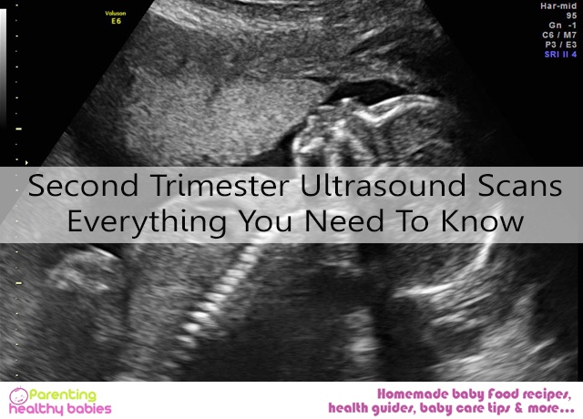
Second Trimester Ultrasounds
Ultrasound in the second trimester is offered between 18th and 22nd week, most commonly in the 20th week. It is used to check the growth and development of the fetus along with the position of placenta in the uterus. The sex of the baby may also be determined after the 20th week. This ultrasound helps rule out many developmental abnormalities and markers for chromosomal disorders in the fetus. Since many measurements and angles need to be covered, the ultrasound takes about 30 to 40 minutes.
Second trimester ultrasounds are primarily abdominal in nature where the health care provider will apply a small quantity of gel on the lower abdomen and see the fetus with the help of a transducer (a small probe that emits sound waves which are then reflected by the fetus and other internal organs).
Wear comfortable, loose two-piece clothing and be sure to ask your health care provider beforehand if you need a full bladder.
Other Names of Second Trimester Ultrasound
- Second trimester anatomy scans
- Fetal anatomy scans
- Targeted imaging for fetal anomalies {TIFFA}
Deciphering Ultrasound
The sonographer will point out the heartbeat and other parts of the body like face and hand. They may be difficult to recognize because the ultrasound imaging is usually done in cross-section. But a simple way to recognize tissue is by their color. Remember, bones will appear white, soft tissues grey and the surrounding amniotic fluid will appear black.
You may want a picture of the baby. Fortunately, most ultrasound centers provide one. The picture is printed on a thermal paper which is heat sensitive. Take care not to laminate it.
Things Checked during UltrasoundScreening
The health care provider would focus on the following during the screening:
- Shape and structure of the head
- Baby’s face to rule out cleft lip
- Baby’s spine
- Baby’s abdominal wall
- Heart rate, rhythm and blood flow
- Kidneys and urine flow to the bladder
- Length of fetus’ arms, legs, hands and feet
- Position of placenta, umbilical cord and amount of amniotic fluid.
Measurements to Look out for
Different measurements are important in different trimesters. While in the first trimester Crown Rump Length (CRL) was of primary importance to determine the expected date of delivery, in the second trimester a few other measurements take up the spot. They are:
- Head Circumference (HC): Head Circumference is one of the important measurements of the second trimester. Not only does it provide an adequate reflection of the health of the fetus, it also helps in ruling out or warn about threatening conditions like neural defects, encephalopathy etc.
- Bi-Parietal Diameter (BPD): bi-parietal diameter is again the measurement of the head. In this case, it is the measure of the length from ear to ear.
Head circumference and bi-parietal diameter are used together to determine expected date of delivery in the second trimester.
- Abdominal Circumference (AC): abdominal circumference provides an accurate measure of the nutritional status of the baby. It reflects the amount of nutrition it receives from the mother and rules out threatening metabolic conditions.
- Femur Length (FL): femur length is the length of the thigh bone of the fetus. It helps to determine the growth rate of the fetus.
Sometimes Humerus Length, nasal bone length, and total cerebral diameters are also measured.
Conditions to Check for
Some rare conditions can be known at this stage. These conditions are rare developmental defects that may be life threatening or debilitating. Others may be easily treatable and manageable. Knowing about these conditions in advance helps to prepare parents for the consequences and may enable them to seek counseling and explore other options.
- Anencephalopathy: absence of top of head.
- Cleft lip and palate
- Exomphalos: defective abdominal wall
- Missing or short limbs
- Spina bifida: defective spinal cord
- Missing or abnormal kidneys
- Hydrocephalous (increased fluid in skull)
- Patau’s syndrome
- Heart defects
In case of heart defects, a fetal echo ultrasound may be advised by your health care provider.
Normal Second Trimester: Week by Week Report
- Week 14: fetus has a CRL of 3 to 4 inches. Genitalia are formed but not clearly visible. Liver and spleen start producing RBCs
- Week 15: CRL is 4 to 4.5 inch. Peculiar hair pattern on scalp can be seen. Bones harden and appear brighter and whiter.
- Week 16: lower limbs are now developed. Toe nails are forming. Eyes and ears take proper shape and location.
- Week 17: Fetal movements begin.
- Week 18: CRL is 5 to 5.5 inches. Distance around the abdomen (abdominal circumference can be measured. Bones and nerve endings related to hearing develop. Sex can be clearly identified if the baby is in the right position.
- Week 20: CRL 5.75 to 6.5 inches. Hair and nails grow. Fetal movements can be seen on the ultrasound screen.
- Week 21: CRL is 7 to 7.5 inches. Baby swallows amniotic fluid regularly. Bone marrow start producing RBCs. Baby freely moves and wriggles around. Heart and lungs can be identified clearly.
- Week 22: Eyebrows form. Sense of taste and smell develops.
- Week 23: CRL is 8 inches. Fat tissue start to form. The fetus begins to gain weight. The baby also starts rapid eye movements. Your baby can now dream!
- Week 24: blood flow can be seen clearly. The fetus reacts to noises with a blink-startle response.
- Week 25: The hearing is now well developed. The baby can now hear and react to the mother’s voice. It will already recognize the mother at the time of birth due to the voice!
- Week 26: eyelashes form. CRL is now 9.5 inches.
- Week 27: liver matures. Immune system starts to develop. The baby has grown so much that it becomes difficult to get a complete profile in the womb.
Week 27 marks the end of the second trimester. Now you have only one more trimester to go. Eat well, stay hydrated and take utmost care of yourself. The baby is happy when the mother is happy!!
Stay informed, stay healthy!!


