
Ultrasound is a sound wave with a frequency higher than 20kHz used to look at organs and structures inside the body. It is same as normal sound in its physical properties, but humans cannot hear it. Ultrasound devices range with frequencies from 20kHz to several gigahertz.
Ultrasound is a type of imaging which is used to view the heart, blood vessels, kidneys, liver and other organs. Doctors use ultrasound to screen the foetus. Ultrasound does not expose you to radiation. It helps to diagnose the causes of pain, swelling and infection in the body’s internal organs and to examine a foetus in pregnant women and the brain and hips in infants. It is also used to guide biopsies, diagnose heart conditions, and assess damage after a heart attack. Ultrasound is safe, non-invasive, and does not use ionizing radiation.
All You Need to Know about Ultrasound Colour
Ultrasound procedure
At the time of testing, you lie on a table and a special technician or doctor moves a device called a transducer over parts of your body. The transducer emits out sound waves which rebound off the tissues inside your body. The transducer also captures the waves that bounce back. This bouncing back or echo gives the ultrasound image its features. The ultrasound machine creates images from the sound waves.
How does it capture an image?
Ultrasound will travel through blood in the heart chamber and if it hits a heart valve, it will rebound back. If there are no gallstones, it will travel straight through the gallbladder, but if there are stones, it will rebound back from them.
Uses
Ultrasound is commonly used for diagnosis, for treatment and for guidance during procedures such as biopsies. An ultrasound scan detects tumour (cancerous, or a fluid-filled cyst). It can diagnose problems with muscles, blood vessels, soft tissues, tendons, and joints. It is used to scrutinize a frozen shoulder, tennis elbow, carpal tunnel syndrome, and others.
Doppler ultrasound can compute the flow of blood in a vessel or blood pressure. As ultrasound turns sound waves into images, it can be used to determine the speed of the blood flow and any obstructions.
An echocardiogram (ECG) is an example of Doppler ultrasound. It is used to create pictures of the cardiovascular system and to measure blood flow and cardiac tissue movement at specific points. It can compute the function and state of the cardiac valve, deformities in the heart, valvular regurgitation, or blood leaking from valves, and it can show how well the heart pumps out blood.
It can also be used to examine the walls of blood vessels, check for deep vein thrombosis or an aneurysm (a condition where blood clots form in veins deep in your body, usually in your legs. Your doctor can use it to check for issues with blood flow, such as clots in your veins or blockages in your arteries), check foetal heart and heartbeat, evaluate for plaque build-up and clots, assess for blockages or narrowing of arteries. It can lead to more serious problems, such as a clot in your lungs. It can be life-threatening. A carotid duplex would reveal how blood cells move through the carotid arteries.
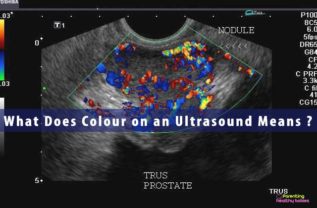
Colours on an Ultrasound
What does red and blue colour denote?
Colour is blue or red depending on whether the blood movement is towards the ultrasound probe or away from it.
Blue represents that the blood flow is away from the probe and red represents that the blood flow is towards the probe.
RED means flow in one direction while BLUE means flow in the opposite direction. Combination of RED & BLUE can be the consequence of circular flow, turbulence, or coherent flow. Different shades of red and blue are used to show velocity. Lighter shades of colour are allocated to higher velocities. Turbulent flow is present when there are large variations in flow velocity within the sample region.
Power Doppler and Colour Doppler Ultrasound
Power Doppler is a form that is more sensitive to slow flow but is described as one color, e.g. gold, orange, and yellow.
Colour Doppler displays the direction and flows velocities of the blood superimposed on the cross-sectional image. In this technology, different pulses are sent across an image line, and the phase shift between the different signals is measured at two sampling points. This phase shift is proportional to the velocity of the reflecting object. A colour is assigned to the direction of flow according to whether it is away from or towards the transducer.


