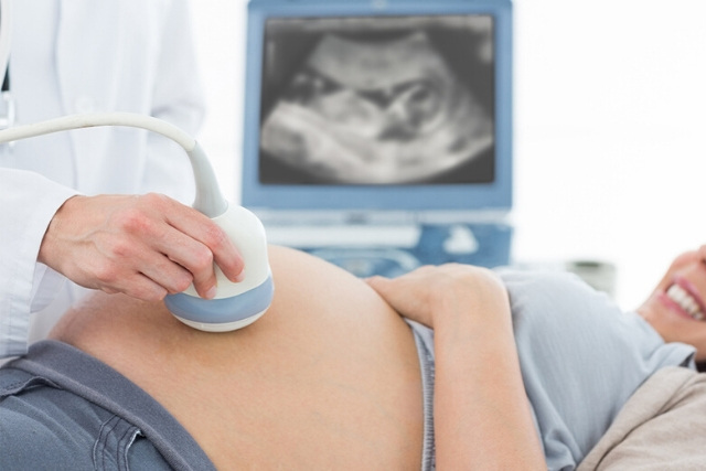
Fetal echocardiogram is used to diagnose cardiac conditions in the fetal stage because cardiac defects are amongst the most common birth defects. So, it is important to diagnosis in the fetal stage as it might help provide an opportunity to plan and manage the baby as and when the baby is born.
In this article:
What is Fetal Echocardiography?
When is Fetal Echocardiography Used?
Is Preparation Needed for the Procedure?
How is a Fetal Echocardiogram Performed?
Risks of Fetal Echocardiography
Result
Is this Test Important?
All You Need to Know about Fetal Echocardiogram
What is Fetal Echocardiography?
Fetal echocardiography is a test similar to an ultrasound test. This exam allows the doctor to better see the structure and also the function of the unborn child’s heart. It’s typically done in the second trimester, between weeks 10 to 24.
The exam uses sound waves that ‘echo’ of the structure of the fetus’s heart. A machine analyzes these sound waves and creates a picture or echocardiogram of the heart’s interior. This image provides information on how the baby’s heart formed and whether it’s working properly. It also enables the doctor to see the blood flow through the fetus’ s heart. This in-depth look allows the doctor to find any abnormalities in the baby’s blood flow or heartbeat.
When is Fetal Echocardiography Used?
Not all pregnant women need a fetal echocardiogram. For most women, a basic ultrasound will show the development of all four chambers of the baby’s heart.
The doctor may recommend that if the procedure is done previously and the tests weren’t conclusive or if detected an abnormal heartbeat in the fetus. This test can also be used if –
- The unborn child is at risk for a heart abnormality or other disorder
- Family history of heart disease
- Given birth to a child with a heart condition
- Used drugs or alcohol during the pregnancy
- Taken certain medications or been exposed to medications that can cause heart defects like epilepsy drugs or prescription acne drugs
- Other medical conditions like rubella, type 1 diabetes, lupus or phenylketonuria
Some doctors perform this test. But usually, an experienced ultrasound technician or ultrasonographer performs the test whereas cardiologist who specializes in pediatric medicine will review the results.
Is Preparation Needed for the Procedure?
No preparation is needed for this test. Unlike other prenatal ultrasounds, no need for full bladder for the test. The test can take anywhere from 30 minutes to two hours to perform.
How is a Fetal Echocardiogram Performed?
This test is similar to a routine pregnancy ultrasound and if it’s performed through the abdomen it is known as abdominal echocardiography whereas if it’s performed through the vagina it is known as transvaginal echocardiography.
Abdominal echocardiography is also similar to an ultrasound. An ultrasound technician first asks to lie down and expose the belly, then they apply a special lubricating jelly to the skin. The jelly prevents friction so that the technician can move an ultrasound transducer which is a device that sends and receives sound waves, over the skin. The jelly also helps transmit sound waves. The transducer sends high-frequency sound waves through the body. The waves echo as they hit a dense object such as the unborn child’s heart. Those echoes are then reflected back into a computer. The sound waves are too high-pitched for the human ear to hear and the technician moves the transducer all around the stomach to get images of different parts of the baby’s heart. After the procedure, the jelly is cleaned off the abdomen and then free to return to normal activities.
Transvaginal echocardiography undress from the waist down and lie on an exam table. A technician will insert a small probe into the vagina. The probe uses sound waves to create an image of the baby’s heart. It is typically used in earlier stages of pregnancy and may provide a clearer image of the fetal heart.
Risks of Fetal Echocardiography
There are no associated risks because it uses ultrasound technology and no radiation.
Result
During the follow-up appointment, the doctor will explain the results and answer the questions. Generally, normal results mean the doctor found no cardiac abnormality.
If the doctor found an issue such as heart defect, rhythm abnormality or other problems, there is a need for more tests such as fetal MRI scan or other high-level ultrasounds. The doctor will also refer to resources or specialists for treating the unborn child’s condition.
May also need to have an echocardiograph done more than once and need additional testing if the doctor thinks something else could be wrong.
It’s important to remember that the doctor can’t use the results of echocardiography to diagnose every condition. Some problems, such as a hole in the heart are difficult to see even with advanced equipment and the doctor will explain what they can and can’t diagnose using the test results.
Is this Test Important?
Abnormal results from fetal echocardiography can be inconclusive or require to get more testing to find what’s wrong. Sometimes problems are ruled out and no further testing is needed. Once the doctor diagnoses a condition, better manage the pregnancy and prepare for delivery.
Conclusion
Results from this test will help and the doctor will plan treatments to the baby may need after delivery such as correctional surgery.
References
https://www.ncbi.nlm.nih.gov/pmc/articles/PMC2747399/
https://www.healthline.com/health/fetal-echocardiography#importance
Hope this article was of help to you! Please share your comments/queries/tips with us and help us create a world full of Happy, Healthy Babies and Empowered Women!!
