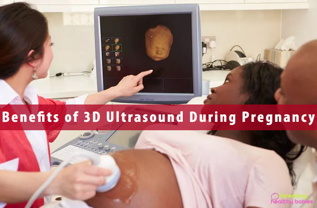
If you are expecting a baby, then one of the most exciting things in your nine months pregnancy period is the sessions of ultrasonography. Getting to hear your baby’s heartbeat for the very first time can be a life-changing experience altogether. The ultrasound gives us a vision into what we are otherwise deprived of, the vision of the baby in the womb. Most of the women are familiar with the traditional 2D process of ultrasonography where only a two-dimensional picture of the foetus can be seen. Ultra-sonographies during pregnancy are something every woman looks up to. If you have been through this, you know exactly what I am talking about!
What is a 3d ultrasound?
A traditional two-dimensional ultrasound works by “hearing” the sound waves which after being directed down, are passed straight back up again. In 3d ultrasounds however, these same sound waves are directed, but for these they are directed down at various and different angles, thus enabling the receiving equipment to “look” at the image as three-dimensional. This new process known as “surface rendering”. The would-be moms therefore get a three-dimensional view of their baby, in a way that they get to perceive the baby much better. In standard 2d scans, usually a blurry grey outline of the foetus is visible, but in the 3d technology, a much clearer structure is available. Nowadays, owing to the latest technology, even 4d sonographies are available!
3D Ultrasound During Pregnancy: 11 Amazing Benefits
Detailed observation
A three-dimensional ultra-sonography provides a far more detailed analysis and observation of the foetus than that provided by a standard two-dimensional ultra-sonography. The clarity and details are relatively much more updated and technical.
Confirmation of gestational age and expected due date
The level of accuracy in 3d ultrasounds are much more than that in the traditional 2d ones. Thus, these are more trustworthy to the doctors as well. Hence, these are used for a confirmation of the gestational age of the foetus and the estimated due date of delivery.
Detecting physical abnormality
The doctor gets a much clearer picture of the foetus from a three-dimensional ultrasound than from a traditional two-dimensional one. This way, it becomes easier for the doctor to detect an abnormality in the foetus, if any. Detection at an early stage of course helps in better treatment in the long run.
Identification of high-risk pregnancies
There are cases where the mother is at high risk due to some complication but that is not detected until delivery. These 3d ultrasounds help detect cases where the pregnancy can be fatal for the mother and helps the doctor take further actions regarding the same. This in turn facilitates the safety of the mother.
Prediction of the child’s gender
In countries where it is legal, most couples do not want to wait for nine whole months to find out the gender of the child. 3d ultrasounds are effective in detecting the gender of the child very early by looking at the chromosomes. While a combination of xx in the chromosome means the female gender, a combination of xy means the male.
Detects behavioural concerns
The specialized 3d ultrasounds are effective in detecting the behavioural concerns of the foetus which further helps in diagnosing problems in the nervous system and brain. About the 3d ultrasound, spain’s instituto bernabeu states that “there is an ability to observe, measure and evaluate the behavior of the foetus and its general movements in a manner which is more comprehensive in comparison to that used in conventional ultrasounds.”
Emotional bonding
Owing to the advanced technology used 3d ultrasounds, parents get to form a special emotional bond with their baby even before he or she is born. They can even observe their movements, making the experience even more memorable.
Detection of cysts, polyps or fibromas
3d ultrasounds help detect the abnormalities in the mother such as cysts, polyps fibromas and so on. At certain cases if these are found malignant later, the matter might go out o hand and it might prove to be fatal for both the baby as well as the mother. Hence, an early detection of its position and condition is always beneficial both for the mother as well as the child.
Saves time
The 3d ultrasounds help save a lot of time, both for the mother as well as for the doctor as they are much more quick and convenient compared to the standard 2d ultrasounds that are usually used.
Measurement of endometrial hyperplasia
Endometrial hyperplasia is a condition governed by higher secretion of estrogen than progesterone. When the process of ovulation stops after conception, excess secretion of estrogen leads to thickening of the endometrial layer which further leads to unnecessary crowding of the cells which can be fatal for the mother and might even lead to cancer. 3d ultrasounds help detect this problem very easily thus saving the mother from a huge life risk.
Better examination of all the criteria
3d ultrasounds are much more sophisticated and technologically advanced that the usual 2d ones. Hence, they are much more effective to examine whether everything is okay and normal with the baby. These include ensuring if there is sufficient amniotic fluid, if the blood low through the placenta to the baby is proper, and if the baby is in the desired head-down position. It is also effective in evaluating the movements of the baby.
There are endless debates about whether the 3D ultrasounds can harm your unborn baby. But let us accept the fact that no product of technological advancement is flawless. Hence, these advantages are reason enough why you should undoubtedly go for a 3D ultrasound during pregnancy if possible!
